3D Imaging & Tomography
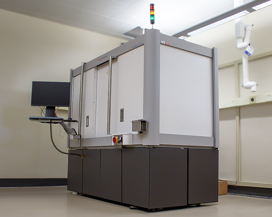
HeliScan microCT Mark II
The HeliScan microCT is a versatile, micro-computed tomography (microCT) imaging system that produces geometrically accurate 3D images of sample materials of any type and shape using conventional circular as well as patented helical trajectories. The HeliScan technology enables continuous scanning of tall samples thus avoiding artefacts commonly associated with stitching. In addition, it produces high quality images over the entire volume of sample imaged for quantitative image analysis. Thermo Fisher Scientific’s scanning technology uses wide X-ray cone angles and a high-flux imaging environment for fast data recording low noise images that accurately represent sample’s microstructure without distortion and are ready for segmentation and analysis.
Features and Specifications
The HeliScan microCT features a water-cooled X-ray source capable of producing an X-ray beam with uniform flux over an extended period; in addition, the instrument maintains X-ray focus during the entire duration of imaging. A key advantage of this design is that it facilitates imaging of tall samples and produces sharper images with uniform intensity throughout the image volume.
The source is integrated with a nitrogen venting line which allows for contaminant-free working conditions for easy filament exchange, and which minimizes downtime.
Key features of the X-ray source include:
- Voltage: 20-160 kV
- Power: 16 W
- Three optimized focus modes: Small, Medium and Large
- Water cooling
- Nitrogen vent line
The HeliScan microCT is equipped with a high-precision, 7 axis motorized motion platform, providing the following movements:
- sample Y-axis: 400 mm travel
- sample Z-axis: 195 mm travel
- sample Rotation: 360° continuous
- sample ROI stages: ±20 mm in X and Y directions
- Load capacity: 15 kg
- Maximum sample diameter: 240 mm
- detector Y axis: 585 mm travel, max. distance 835 mm from source
- detector X axis: ± 50 mm
The HeliScan microCT is equipped with a large area amorphous silicon flat panel detector.
The features of flat panel detector include:
- 3072 × 3072 pixels
- 16 bit
- 800 nm*
- Nominal resolution: 170 nm**
* (2D) Spatial resolution (or 10% MTF resolution) – Resolution is the distance between two objects (or cavities) at which they can still be identified independently.
** Nominal Resolution – Voxel size at the highest possible magnification.
 HeliScan microCT Mark II in Chemistry & Materials Building, Room 168
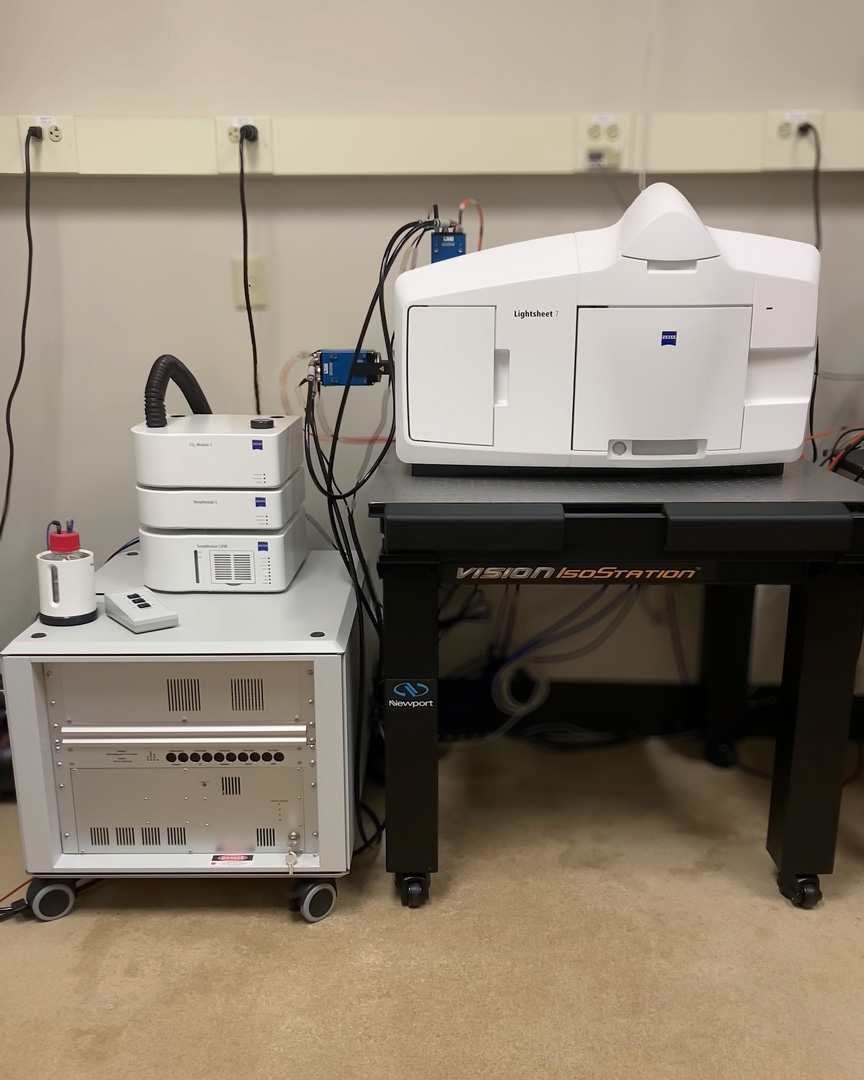
Zeiss Light Sheet 7
Light Sheet Fluorescence Microscopy (LSFM) is an extremely powerful alternative to established fluorescence imaging techniques, especially for 3-D imaging within whole live organisms and large tissue explants. Ideal for fast and gentle imaging of whole living model organisms, tissues, and cells as they develop – over extended periods of time capable of imaging large optically cleared specimens in toto – with subcellular resolution.
By selectively illuminating the observed optical section with a thin sheet of light, photo bleaching is reduced to a minimum, making light sheet microscopy ideal for nondestructive imaging of fragile samples over extended periods of time. Equipped with an incubation system with temperature and CO2 control for long-term in vivo experiments.
Features and Specifications
Magnifi-cation |
Objective Type |
NA |
Immer-sion |
WD (mm) |
|---|---|---|---|---|
| 2.5x | Fluar | 0.12 | Clearing | 8.7 |
| 5x | EC Plan-Neoflua | 0.16 foc | Water/Clearing | 18.5 |
| 5x | EC Plan-Neofluar | 0.16 | Clearing | 18.5 |
| 10x | W Plan-Apochromat | 0.50 | Water | 3.7 |
| 20x | W Plan-Apochromat | 1.00 | Water | 2.4 |
| 20x | W Plan-Apochromat | 1.00 | Clearing | 6.4 |
405nm, 445nm, 488nm, 514nm, 561nm, 638nm
Two liquid cooled sCMOS cameras, pco.EDGE
Fluoro-chromes |
Mirror |
Filter 1 (nm) |
Filter 2 (nm) |
|---|---|---|---|
| DAPI/GFP | SBS LP 490 | BP 420-470 (DAPI) | BP 505-545 (GFP) |
| DAPI/RFP | SBS LP 510 | BP 420-470 (DAPI) | BP 505-545 (GFP) |
| GFP/mCherry | SBS LP 560 | BP 505-545 (GFP) | LP 585 (mCherry) |
| GFP/ Draq5(toto3) | SBS LP 560 | BP 505-545 (GFP) | LP 660 (Draq5(toto3)) |
| RFP/ Draq5(toto3) | SBS LP 640 | BP 575-615 (RFP) | LP 660 (Draq5(toto3)) |
Color |
Diameter (mm) |
|---|---|
| Red | 0.68 |
| Black | 1.00 |
| Green | 1.50 |
| Blue | 2.15 |
 Zeiss Light Sheet 7 in Chemistry & Materials Building, Room 155