Crystallographic Analysis
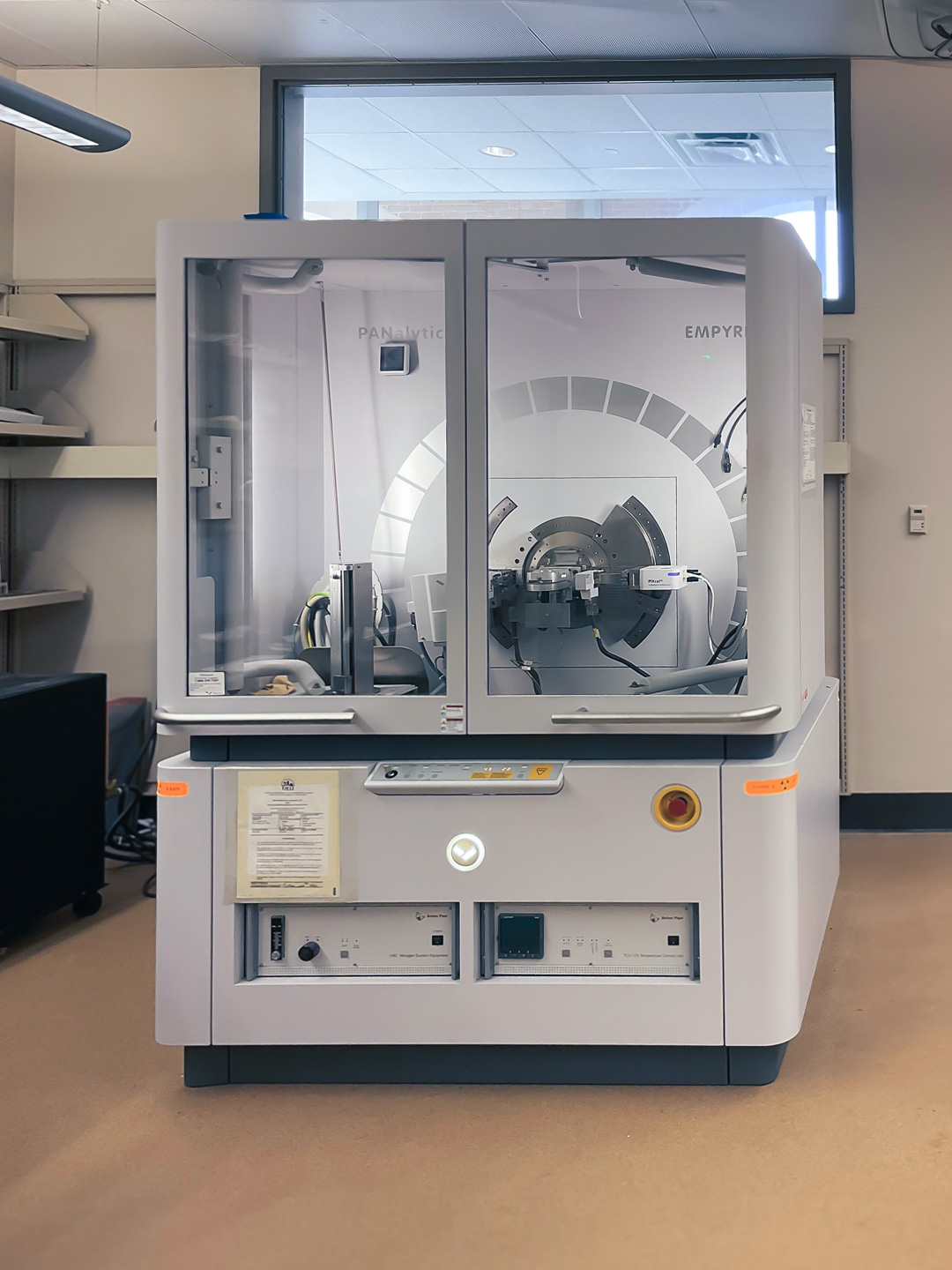
PANalytical Empyrean X-Ray Diffractometer
This PANalytical Empyrean multipurpose diffractometer is equipped with PreFIX (pre-aligned, fast interchangeable X-ray) modules, making a change in the optical path time efficient and user friendly. It allows applications to be done in various ways: from ‘simple’, by just adding a few slits and setting the analysis software to a standard geometry, to top performance using dedicated optics, sources and detectors.
With the PIXcel3D X-ray detector, which is based on Medipix3 technology, PANalytical has been able to design a multipurpose system that handles measurements on powders, thin films, nanomaterials and bulk objects in one instrument, with no compromise in data quality. The PreFIX mounting method of optics allows users to switch between different applications in a matter of minutes.
- Maximum usable range: -111° < 2θ < 168°
- 2θ linearity equal or better than ±0.01°
- Temperature range of -150°C to 400°C
- Capillary tube can be used for air sensitive materials or small sample size
- Grazing Incidence X-Ray Diffraction* (GIXRD) provides surface information or depth profiling on randomly oriented polycrystalline materials.
*GIXRD can help to distinguish thin film signals from the substrate or other layers, measure residual stress as a function of depth, and perform phase ID analysis at the surface and as a function of depth.
Electron Backscatter Diffraction (EBSD) with the Oxford Instruments Symmetry system on our Xe P-FIB/SEM
EBSD can provide valuable information about the grain structure, texture, and crystallographic phases of a sample (e.g. metals, semiconductors, ceramics).
In general, a carefully prepared, plan sample is scanned by a focused electron beam. Electrons interacting by Bragg diffraction within the top nano meters of the surface form a diffraction pattern (Kikuchi pattern) at every scan point which can be detected and analyzed. The resulting maps can cover grains of millimeter, micrometer down to the nanometer scale, depending on the scan resolution used.
With the same system but in a different geometry, electron transparent samples like TEM lamellae can be analyzed in transmission mode. This technique is called Transmission Kikuchi Diffraction (TKD).
Insert image/photo here.
